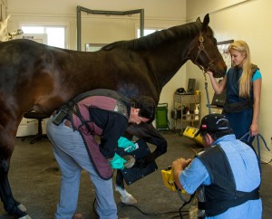
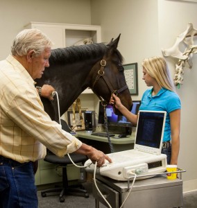
Early diagnosis and treatment is key to treating any disease. At Specifically Equine Veterinary Service, we offer multiple diagnostic options. Our capabilities include endoscopy, gastroscopy, digital radiography, digital ultrasonography and MRI. Our digital equipment allows us to forward images electronically to referral doctors for updates and specialists for timely second opinions.
Gastroscopy and Endoscopy
Endoscopy and gastroscopy allow a veterinarian to see areas such as the nasal passage, pharynx, trachea, esophagus, stomach, urinary tract, uterus and bladder. Using our one meter Endoscope we are able to visualize the upper respiratory system including the sinuses, guttural pouches and larynx. This allows for early recognition and treatment of many respiratory diseases. The 3 meter gastroscope allows us to visualize the stomach, pylorus and the upper most portion of the small intestine. We are then able to detect stomach abnormalities such as ulcers.

Digital Radiography
Digital radiology allows instant high detail radiographs, providing information that often was not visualized on plain film x-rays. Subtle lesions can be examined from the level of individual hairs down to deep bone.Under high magnification, images remain very clear and margins retain sharp contrast. The “computerized” capability of this system allows us to adjust tones to best match the diagnostic needs of the individual horse and to detect very small problems which are not as visible on traditional x-rays.

Digital Ultrasound
Digital ultrasound allows for real-time viewing and evaluation of soft tissues, such as tendons and ligaments, to identify injuries, provide a prognosis and evaluate the progress of healing. Deep structures such as the pelvic sacro-iliac joints and cervical facets (neck joints) can also be examined for signs of arthritis. Digital ultrasound images are often used to guide therapeutic injections of these areas as well.
MRI
Magnetic Resonance Imaging uses a strong magnetic field and radio waves to create detailed images of the horse’s anatomy. MRI is the typically the next step when traditional methods such as radiographs and ultrasound have failed to identify the cause of the problem. The MRI is performed under general anesthesia to allow for proper positioning and minimize movement during the exam. With orthopedic conditions of the extremities, both limbs are imaged at the respective sites of injury for a complete evaluation and comparison between limbs. Specifically Equine partners with mobile imaging specialists MREquine to perform testing on site at the clinic.
